



- 商品介绍
- 规格参数
- 包装参数
General description
Atlastin-1 (UniProt Q8WXF7; also known as Brain-specific GTP-binding protein, GBP-3, GTP-binding protein 3, Guanine nucleotide-binding protein 3, hGBP3, Spastic paraplegia 3 protein A) is encoded by the ATL1 (also known as GBP3, HSN1D, SPG3, SPG3A) gene (Gene ID 51062) in human. Atlastin-1 is a dynamin/Mx/guanylate-binding protein superfamily member with three conserved guanylate-binding/GTPase active site motifs, P-loop (a.a. 74-81), DxxG (a.a. 146-149), and RD (217-218). Atlastin-1 is a multimeric integral membrane protein localized on ER and cis-Golgi apparatus and widely expressed in many tissues, including the smooth muscle, adrenal gland, kidney, testis, lung, and brain. Atlastin-1 interacts with the microtubule-severing ATPase spastin as well as with the DP1/Yop1p and reticulon families of ER-shaping proteins, Atlastin-1 knockdown results in decreased number of neuronal processes and impaired axon formation in cultured cortical neurons, while atlastin-1 overexpression increases total dendrite length both in vivo and in vitro. ALT1 gene mutations cause abnormal ER morphology and are linked to hereditary spastic paraplegias (HSPs), a genetically heterogeneous group of neurological disorders, includiing SPG3 (spastic paraplegia 3, autosomal dominan) and HSN1D (neuropathy, hereditary sensory, 1D), characterized by lower limb spasticity and weakness. Human atlastin-1 is a 492-amino acid protein that passes through the ER membrane twice (a.a. 450-470, 472-492), having both its N- and C-terminal ends at the cytoplasmic side (a.a. 1-449, 493-558).
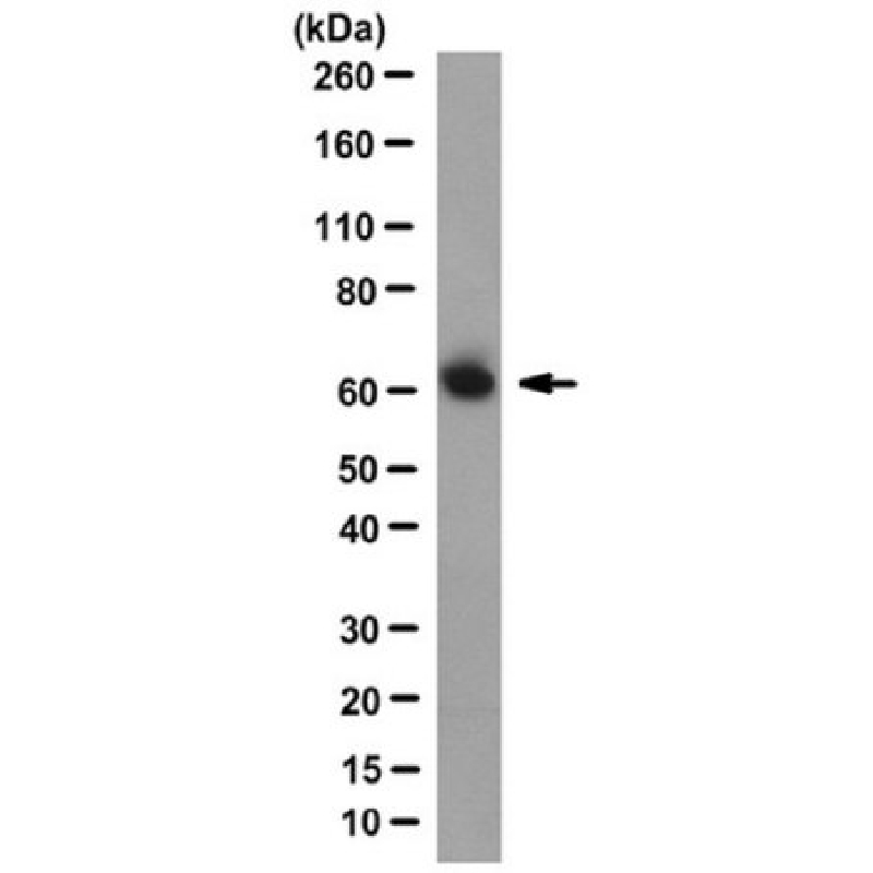
~63 kDa observed. 63.54/63.06 (human isoform 1/2), 63.52 kDa (monkey), and 63.38 kDa (mouse and rat) calculated.
Immunogen
Linear peptide corresponding to the N-terminal sequence of human Atlastin-1.
Application
Anti-Atlastin-1 Antibody, clone 3194 is an antibody against Atlastin-1 for use in Western Blotting, Immunocytochemistry.
Western Blotting Analysis: 10 µg/mL from a representative lot detected Atlastin-1 in 10 µg of mouse brain tissue lysate.
Immunocytochemistry Analysis: A representative lot localized Atlastin-1 immunoreactivity with that of REEP1 by dual fluorescent immunocytochemistry staining of paraformaldehyde-fixed rat cortical neurons (Park, S.H., et al. (2010). J. Clin. Invest. 120(4):1097-1110).
Immunocytochemistry Analysis: A representative lot localized overexpressed human Atlastin-1 in paraformaldehyde-fixed COS7 transfectants by fluorescent immunocytochemistry (Park, S.H., et al. (2010). J. Clin. Invest. 120(4):1097-1110).
Biochem/physiol Actions
Clone 3194 targets an N-terminal epitope present in both spliced isoforms of human Atlastin-1 reported by UniProt (Q8WXF7).
Physical form
Format: Purified
Analysis Note
Evaluated by Western Blotting in human brain tissue lysate.
Western Blotting Analysis: 2.0 µg/mL of this antibody detected Atlastin-1 in 10 µg of human brain tissue lysate.
Other Notes
Concentration: Please refer to lot specific datasheet.
| biological source | mouse |
| Quality Level | 100 |
| conjugate | unconjugated |
| antibody form | purified immunoglobulin |
| antibody product type | primary antibodies |
| clone | 3194, monoclonal |
| species reactivity | mouse, rat, human |
| species reactivity (predicted by homology) | monkey (based on 100% sequence homology) |
| technique(s) | immunocytochemistry: suitable,western blot: suitable |
| isotype | IgG2aκ |
| NCBI accession no. | NP_056999 |
| UniProt accession no. | Q8WXF7 |
| target post-translational modification | unmodified |
| Gene Information | human ... ATL1(51062) |
| 长度(mm) | |
| 宽度(mm) | |
| 高度(mm) | |
| 重量(kg) |
-
询价
-
¥ 2622.09
-
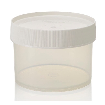
¥ 2290.21
-
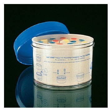
¥ 1259.40
-
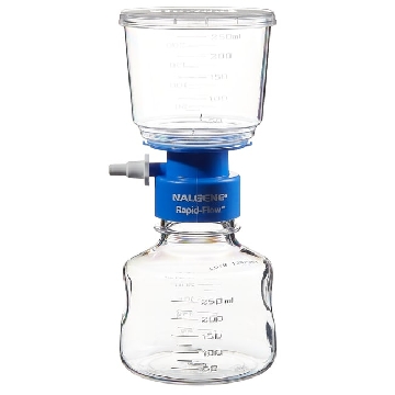
¥ 1866.29
-

¥ 5887.85
-
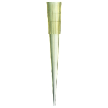
¥ 238.17
-
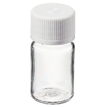
¥ 5167.58
-
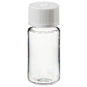
¥ 5725.45
-

¥ 4390.38
-
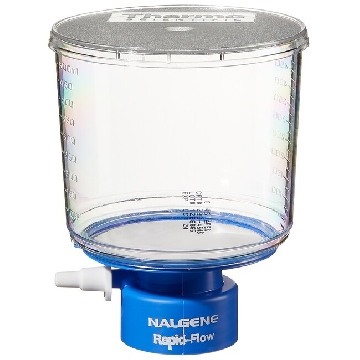 瓶顶过滤器-500ml容量,MF75TM系列,聚苯乙烯外壳,75mmPES滤膜,孔径0.45um,适合瓶颈尺寸33mm,12/箱,295-3345,Nalgene,Thermofisher,赛默飞世尔
¥ 1938.54
-
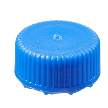
¥ 2842.54
-
¥ 2666.57
-
¥ 1476.00
-
¥ 1749.15
-

¥ 9909.51
-
¥ 2888.88
-
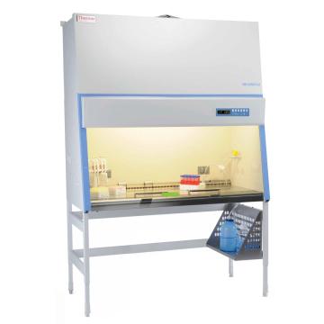
询价
-
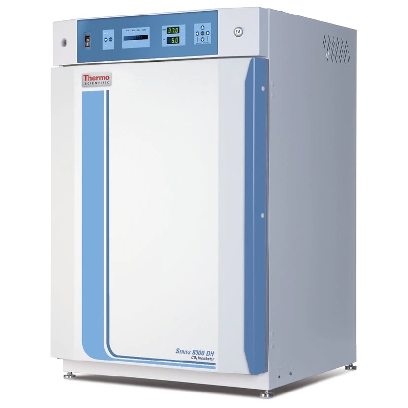
¥ 44224.19
-
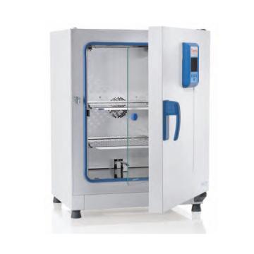
¥ 37675.92









 微信小程序
7X24小时在线咨询
微信小程序
7X24小时在线咨询