





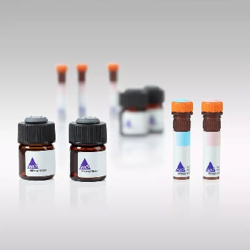
- 商品介绍
- 规格参数
- 包装参数
Immunogen
Lung tumor cell lines A549 and A2182.
Application
Immunohistochemistry: 10-20 μg/mL Optimal working dilutions must be determined by end user.
Tumor Characterization
For tumor characterization a two-step experimental assay is recommended:
1. Characterization of the tissue origin of a tumor with the aid of anti-cytokeratin pan, anti-desmin, anti-glial fibrillary acidic protein, anti-neurofilament and anti-vimentin.
2. Characterization of an epithelial tumor using antibodies to cytokeratin classes which are characteristic for different tissues, e.g. anti-cytokeratin No. 7, anti-cytokeratin No. 18. Sample material: Normal tissue, tumor tissue following surgery or up to 24 h after autopsy, frozen sections or formaldehyde-fixed paraffin embedded sections,
Detection method
Immunohistochemical analysis (detection of the primary antibody e. g. by anti-mouse Ig-peroxidase or anti-mouse Ig-FITC).
Procedure:
Ideal frozen sections are obtained from shock-frozen tissue samples. The frozen sections are air-dried and then fixed with acetone for 5-10 min at -15 to -25°C. Excess acetone is allowed to evaporate at 15-25°C. Material fixed in alcohol can also be used.
Cytocentrifuge preparations of single cells or cell smears are also fixed in acetone. These preparations should, however, not be air dried, the excess acetone is removed by briefly washing in phosphate-buffered saline (PBS). Further treatment as follows:
• Overlay the preparation with 10 - 20 μL antibody solution and incubate in a humid chamber for 30 - 60 min at 15-25°C.
• Dip the slide briefly in PBS and then wash in PBS 3 times for 3 min each (using a fresh PBS bath in each case).
• Wipe the margins of the preparation dry, overlay the preparation with 10 - 20 μL of a solution of anti-mouse lg-FITC or anti-mouse Ig-peroxidase and allow to incubate in a humid chamber for 30 min at 15-25°C.
• Wash the slide in PBS as described above.
The preparation must not be allowed to dry out during any of the steps. lf using an indirect immunofluorescence technique, the preparation should be overlaid with a suitable embedding medium (e. g. Mowiol, Hoechst) and examined under the fluorescence microscope. lf a POD-conjugate is used as the secondary antibody, the preparation should be overlaid with a substrate solution (see below) and incubated at 15-25°C until a clearly visible redbrown color develops. A negative control (e.g. only the labeled secondary antibody) should remain unchanged in color during this incubation period.
Subsequently, the substrate is washed off with PBS and the preparation is stained with hemalum stain for about 1 min. The hemalum solution is washed off with PBS, the preparation is embedded and examined.
Substrate solutions:
Aminoethyl-carbazole: Dissolve 2 mg 3-amino-9-ethylcarbazole with 1.2 mL dimethylsulfoxide and add 28.8 mL Tris-HCI, 0.05 M; pH 7.3; and 20 μL 3% H 2 O 2 , (w/v). Prepare solution freshly each day. Diaminobenzidine: Dissolve 25 mg 3, 3′-diaminobenzidine with 50 mL Tris-HCI, 0.05 M; pH 7.3; and add 40 μL 3% H2O2 , (w/v). Prepare solution freshly each day. Note The antibody solutions have to be absolutely free of precipitate! lf necessary, centrifuge the solutions at high speed prior to use.
This Anti-Cytokeratin Pan Antibody, clone Lu5 is validated for use in IH for the detection of Cytokeratin Pan.
Biochem/physiol Actions
The antibody reacts with an epitope which is common to all cytokeratins respectively with a cytokeratin-associated protein. The antigen recognized by anti-cytokeratin pan is present in all types of epithelia. The antibody reacts with epithelial and mesothelial cells. Exception: Secretory cells are not stained and acinus cells, for example, show only weak staining. The antibody does not react with connective tissue, blood or lymphatic vessels, smooth or skeletal muscles. Exception: When higher antibody concentrations are used, weakly positive reactions can be observed with the smooth muscles of the myometrium, the prostata as well as the gastric muscularis mucosa.The antibodies can be used for differential diagnosis of epithelial tumors, distinction from mesenchymal, neural tumors as well as large-cell lymphomas. Anti-cytokeratin pan stains all epithelial tumors (primary tumors as well as metastases). Exception: Only partially positive staining is observed with theca cell tumors, granulosa cell tumors, adrenal cortex tumors. In mixed tumors (e.g. carcinosarkoma, thymoma, adenolymphoma) only the epithelial regions are stained. Tumors of nonepithelial origin do not react with the antibody. Exception: With high antibody concentrations a weak staining of leiomyosarcoma of the uterus is observed.
Physical form
Format: Purified
Liquid. Buffer = 0.02 M Phosphate buffer, 0.25M NaCl with 0.1% sodium azide.
Preparation Note
The lyophilized antibody is stabe when stored at 2-8°C.The reconstituted antibody solution is stable at 2-8°C for three monthes. The solution can be stored in aliquots at -15 to -25 °C for 12 monthes. Avoid repeat freeze/thaw cycles.
Other Notes
Concentration: Please refer to the Certificate of Analysis for the lot-specific concentration.
Legal Information
CHEMICON is a registered trademark of Merck KGaA, Darmstadt, Germany
| biological source | mouse |
| Quality Segment | 100,300 |
| antibody form | purified antibody |
| antibody product type | primary antibodies |
| clone | Lu5, monoclonal |
| species reactivity (predicted by homology) | mammals |
| manufacturer/tradename | Chemicon® |
| technique(s) | immunohistochemistry: suitable |
| isotype | IgG1 |
| shipped in | wet ice |
| 长度(mm) | |
| 宽度(mm) | |
| 高度(mm) | |
| 重量(kg) |
-
¥ 1259.40
-
¥ 1573.49
-
询价
-
询价
-
¥ 1302.89
-
¥ 9207.92
-
¥ 4173.75
-
询价
-
询价
-
询价
-
询价
-
询价
-
询价
-
询价
-
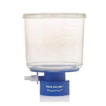
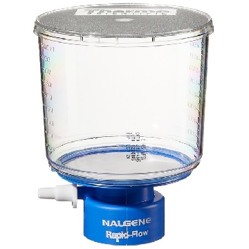 瓶顶过滤器-500ml容量,MF75TM系列,聚苯乙烯外壳,75mmPES滤膜,孔径0.45um,适合瓶颈尺寸33mm,12/箱,295-3345,Nalgene,Thermofisher,赛默飞世尔
¥ 1938.54
-
询价
-
¥ 1564.00
-
¥ 2132.12
-
¥ 2173.94
-
¥ 17652.29









 微信小程序
7X24小时在线咨询
微信小程序
7X24小时在线咨询