


- 商品介绍
- 规格参数
- 包装参数
|
规格 |
|
| Detection Location: | In-Gel Detection |
| Detection Method: | Fluorescent |
| Label or Dye: | Proprietary Mix |
| Product Line: | Krypton™ |
| Target Molecule: | Protein |
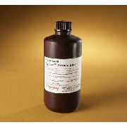
| Quantity: | 500 mL |
|
储存 |
|
| Upon receipt store at 4°C. |
|
描述 |
Thermo Scientific Krypton Fluorescent Protein Stain enables sensitive visualization with fluorescence imagers (520nm excitation; 580nm emission) of proteins separated in polyacrylamide gels or transferred to membranes.
Features of Krypton Fluorescent Protein Stain:
• Intense fluorescent stain—excitation and emission maxima of 520nm and 580nm, respectively (compatible with 532nm excitation light sources and 580 to 600nm emission filters)
• Instrument-compatible—effective with popular laser-based and filter-based fluorescence imagers
• Broad dynamic range—linear quantitative range spans three to four orders of magnitude Sensitive—detect as little as 0.25ng protein with the standard protocol (approx. 2.5 hours); at least as sensitive as fluorescent stains from other suppliers
• Fast—standard (2.5 hours) and rapid (30 minutes) protocols are significantly faster than procedures of other popular fluorescent stains
• Versatile—compatible with proteomics workflows involving downstream mass spectrometry
• Compact—supplied as a 10X solution to save storage space; just dilute with water before use
This fluorescent protein gel stain is for bright yellow-orange fluorescent detection of proteins separated by 1D or 2D polyacrylamide gel electrophoresis (SDS-PAGE). Krypton Stain uses a protein-binding dye with fluorescence properties (Ex/Em = 520/580nm) suitable for visualization with a variety of standard laser-based and filter-based fluorescence imagers and documentation systems. Proteins are detectable at concentrations greater than 0.25ng per band in a typical mini-gel lane (greater than 2ng for the rapid staining protocol). Krypton Stain provides high signal intensity, exhibits minimal protein-to-protein variation, has an excellent quantitative dynamic range and is compatible with subsequent destaining and analysis by mass spectrometry.
For best results, detect bands using visible laser-based imagers equipped with a 532nm laser light source. Although 580nm-filters are optimal for emission, 600nm-filters are also compatible. Stained gels can be imaged on any platform with the respective excitation and emission filters.
| 长度(mm) | |
| 宽度(mm) | |
| 高度(mm) | |
| 重量(kg) |
-
;白色高密度聚乙烯螺旋盖,250ml容量")
询价
-

询价
-

询价
-

询价
-

询价
-

询价
-

询价
-

询价
-
询价
-

询价
-

询价
-
询价
-
,24/箱,867013-0245,Nalgene,Thermofisher,赛默飞世尔")
询价
-

询价
-

询价
-

¥ 145427.01
-

询价
-

询价
-
材质,,带盖,灭菌,单个包装,1个/包/24包/箱,PLT,24WL,FB,ULA,IND,W/LID,S,1/24,型号3473,Corning,康宁")
询价
-

询价


 微信小程序
7X24小时在线咨询
微信小程序
7X24小时在线咨询