




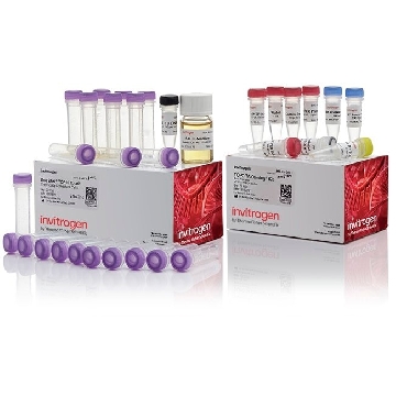
- 商品介绍
- 规格参数
- 包装参数
General description
Fibronectin (FN), or in earlier literature also known as Cold-insoluble globulin (CIG), and is encoded by the gene named FN1/FN is an extracellular matrix protein. Fibronectins bind cell surfaces and various compounds including collagen, fibrin, heparin, DNA, and actin. Fibronectins are involved in cell adhesion, cell motility, opsonization, wound healing, and maintenance of cell shape. Fibronectin also plays an important role in bone development and bone structure formation. Additionally, when fibronectin binds the protein anastellin it forms a multiprotein complex termed superfibronectin that has been shown to inhibit tumor growth, angiogenesis and metastasis. Fibronectin protein can be found extracellularly as well as intracellularly. There are multiple isoforms produced by alternative splicing. Plasma fibronectin (a soluble dimeric form) is secreted by hepatocytes into the blood. Cellular fibronectin (dimeric or cross-linked multimeric forms), are made by fibroblasts, epithelial and other cell types, and is deposited as fibrils in the extracellular matrix.
~260 kDa observed
Immunogen
Epitope: This antibody recognizes the cell attachment fragment of Fibronectin.
Purified human Fibronectin, which contains the Cell Attachment Fragment, from amnion liquid.
Application
Research Category
Cell Structure
Research Sub Category
ECM Proteins
This Anti-Fibronectin (Cell Attachment Fragment) Antibody, clone 3E3, Ascites & Azide Free is validated for use in western blotting, IHC, ELISA, ICC & inhibition for the detection of Fibronectin.
Western Blotting Analysis: 0.5 µg/mL of this antibody detected Fibronectin (Cell Attachment Fragment) in 10 µg of U-251 cell lysate.
Immunohistochemistry Analysis: A 1:500 dilution from a representative lot detected Fibronectin (Cell Attachment Fragment) in human placenta and liver tissues.
ELISA Analysis: A representative lot detected the conformation of Fibronectin adsorbed onto self-assembled monolayers (SAMs) of alkanethiols over a range of Fibroectin surface densities (Kesolowsky, B. G., et al. (2003). J Biomed Mater Res A. 66(2):247-259.).
Immuncytochemistry Analysis: A representative lot detected Fibronectin (Cell Attachment Fragment) in Extracellular matrix deposited by HUVECs (Orecchia, A., et al. (2003). J Cell Sci. 116(Pt 17):3479-3489.).
Inhibition Analysis: A representative lot blocked cell-adhesive epitopes of Fibronectin (Cell Attachment Fragment) (Miller, D. C., et al. (2005). J Biomed Mater Res A. 73(4):476-84.).
Inhibition Analysis: A representative lot blocked RGD-dependent cell adhesion (Tsang, T. M., et al. (2012). J Biol Chem. 287(20):16759-16767.).
Inhibition Analysis: A representative lot inhibited the cell-adhesive epitopes of Fibronectin (Miller, D. C., et al. (2005). J Biomed Mater Res. 73(4):476-484.)
Physical form
0.01M Phosphate Buffer containing no preservative, pH 7.1
Format: Purified
Protein G Purified
Preparation Note
Stable for 1 year at -20°C from date of receipt.
Handling Recommendations: Upon receipt and prior to removing the cap, centrifuge the vial and gently mix the solution. Aliquot into microcentrifuge tubes and store at -20°C. Avoid repeated freeze/thaw cycles, which may damage IgG and affect product performance.
Analysis Note
Evaluated by Western Blotting in HepG2 cell lysate.
Western Blotting Analysis: 0.5 µg/mL of this antibody detected Fibronectin (Cell Attachment Fragment) in 10 µg of HepG2 cell lysate.
Other Notes
Concentration: Please refer to the Certificate of Analysis for the lot-specific concentration.
Disclaimer
Unless otherwise stated in our catalog or other company documentation accompanying the product(s), our products are intended for research use only and are not to be used for any other purpose, which includes but is not limited to, unauthorized commercial uses, in vitro diagnostic uses, ex vivo or in vivo therapeutic uses or any type of consumption or application to humans or animals.
| biological source | mouse |
| Quality Segment | 100 |
| antibody form | purified antibody |
| antibody product type | primary antibodies |
| clone | 3E3, monoclonal |
| species reactivity | human |
| technique(s) | ELISA: suitable,immunocytochemistry: suitable,immunohistochemistry: suitable,inhibition assay: suitable,western blot: suitable |
| isotype | IgG1κ |
| NCBI accession no. | NP_002017 |
| UniProt accession no. | P02751 |
| shipped in | wet ice |
| target post-translational modification | unmodified |
| Gene Information | human ... FN1(2335) |
| 长度(mm) | |
| 宽度(mm) | |
| 高度(mm) | |
| 重量(kg) |
-
询价
-
¥ 1048.84
-
¥ 673.51
-
询价
-
询价
-
询价
-
询价
-
询价
-
询价
-
¥ 1987.51
-
¥ 2067.03
-
¥ 2532.00
-
¥ 661.45
-

询价
-
¥ 1761.75
-
¥ 860.58
-
¥ 1137.02
-
¥ 1066.63
-
¥ 633.78
-
¥ 699.66









 微信小程序
7X24小时在线咨询