



- 商品介绍
- 规格参数
- 包装参数
General description
Cytokeratin 3, also known as 65kDa Cytokeratin or Keratin K3 is a specific keratin found primarily in the corneal epithelium and minorly in other epithelium like that of the esophagus. The corneal epithelium tissue forms the outermost layer of the cornea, which is the clear front covering of the eye. Keratins are divided into two primary types based upon their isoelectric mobility and size, acidic, or type 1:40-55kDa, and basic to neutral or type 2, 56-70kDa. Cytokeratin 3 is a type II cytokeratin. Type II cytokeratins form pairs with other cytokeratins. Cytokeratin 3 is expressed in the corneal epithelium with Cytokeratin 12. Together Cytokeratin 3 and Cytokeratin 12 form molecules known as intermediate filaments. These filaments assemble into strong networks that provide strength and resilience to the corneal epithelium. Mutations in Cytokeratin 3 have been associated with Meesmann′s Corneal Dystrophy a disease characterized by fragility of the anterior corneal epithelium.
~60 kDa observed
Immunogen
Whole cells corresponding to Rabbit Keratin, type II cytosleletal 3.
Application
Immunohistochemistry Analysis: A 1:1000 dilution from a representative lot detected Keratin K3/K76 in bovine cornea epithelial cells.
Immunohistochemistry Analysis: A representative lot was used by an independent laboratory in Keratin 3 expressed epithelial cells on the surface of submerged and AL explants, indicating that they were derived from limbal progenitor cells via migration. (Kawakita, T., et al. (2005) Am J Pathol, 167:381-93.).
Western Blotting Analysis: A representative lot was used by an independent laboratory in Rabbit epithelial Keratin lysate. (Schermer, A, et al. (1986) J. Cell Biol., 103: 49-62).
Western Blotting Analysis: A representative lot was used by an independent laboratory in rabbit corneal tissue lysate. (Cooper, D., et al. (1986) J. Biol. Chem., 261: 4646-54).
Immunocytochemistry Analysis: A representative lot was used by an independent laboratory in human limbal stem cells cultured in medium supplemented with human EGF. (Sharifi, A.M., et al. (2010) Biocell. 34(1):53-55).
Immunofluorescence Analysis: A representative lot was used by an independent laboratory in rabbit corneal and limbal tissues, limbal explant, and epithelial outgrowth on human AM. (Wang D.Y., et al. (2003) Invest Ophthalmol Vis Sci. 44(11):4698-704).
Immunofluorescence Analysis: A representative lot was used by an independent laboratory inRabbit corneal epithelial Keratin cells. (Schermer, A, et al. (1986) J. Cell Biol., 103: 49-62).
Research Category
Cell Structure
Research Sub Category
Cytokeratins
This Anti-Keratin K3/K76 Antibody, clone AE5 is validated for use in Western Blotting and Immunohistochemistry and Immunofluorescence for the detection of Keratin K3/K76.
Physical form
Format: Purified
Protein G Purified
Purified mouse monoclonal IgG1κ in buffer containing 0.1 M Tris-Glycine (pH 7.4), 150 mM NaCl with 0.05% sodium azide.
Preparation Note
Stable for 1 year at 2-8°C from date of receipt.
Analysis Note
Evaluated by Western Blotting in Rabbit eye (cornea) cell lysate.
Western Blotting Analysis: 0.05 µg/mL of this antibody detected Keratin K3/K76 in 10 µg of Rabbit eye (cornea) cell lysate.
Other Notes
Concentration: Please refer to lot specific datasheet.
Replaces: CBL218
Disclaimer
Unless otherwise stated in our catalog or other company documentation accompanying the product(s), our products are intended for research use only and are not to be used for any other purpose, which includes but is not limited to, unauthorized commercial uses, in vitro diagnostic uses, ex vivo or in vivo therapeutic uses or any type of consumption or application to humans or animals.
| biological source | mouse |
| Quality Segment | 100 |
| conjugate | unconjugated |
| antibody form | purified immunoglobulin |
| antibody product type | primary antibodies |
| clone | AE5, monoclonal |
| species reactivity | rabbit, bovine |
| technique(s) | immunofluorescence: suitable,immunohistochemistry: suitable,western blot: suitable |
| isotype | IgG1κ |
| NCBI accession no. | XP_002711051 |
| UniProt accession no. | Q29426 |
| shipped in | wet ice |
| target post-translational modification | unmodified |
| Gene Information | human ... KRT3(3850) |
| 长度(mm) | |
| 宽度(mm) | |
| 高度(mm) | |
| 重量(kg) |
-
询价
-
询价
-
¥ 1596.77
-
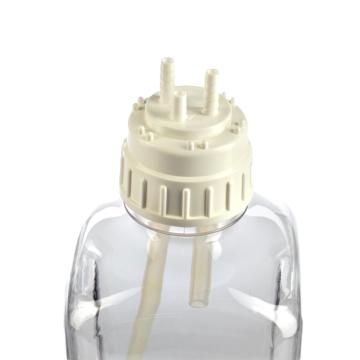
询价
-
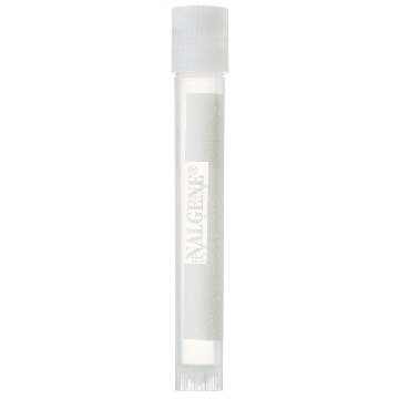
¥ 1683.78
-
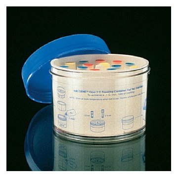
¥ 1259.40
-

¥ 5887.85
-
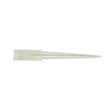
询价
-
询价
-
询价
-
询价
-

询价
-
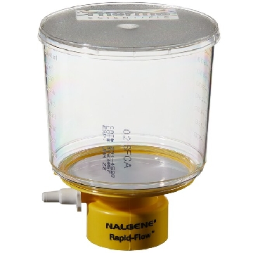 瓶顶过滤器-500ml容量,MF75TM系列,聚苯乙烯外壳,75mmSFCA滤膜,孔径0.2um,适合瓶颈尺寸45mm,12/箱,291-4520,Nalgene,Thermofisher,赛默飞世尔
¥ 2856.42
-
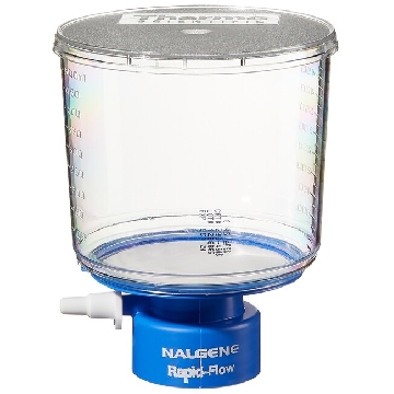 瓶顶过滤器-500ml容量,MF75TM系列,聚苯乙烯外壳,75mmPES滤膜,孔径0.45um,适合瓶颈尺寸33mm,12/箱,295-3345,Nalgene,Thermofisher,赛默飞世尔
¥ 1938.54
-
¥ 2617.87
-
¥ 2173.94
-
¥ 5990.26
-
¥ 3662.39
-
询价
-
询价









 微信小程序
7X24小时在线咨询
微信小程序
7X24小时在线咨询