



- 商品介绍
- 规格参数
- 包装参数
General description
Metavinculin (MV) and its a.a. 916-983 truncated splice variant Vinculin (UniProt P18206) are cytoskeleton proteins encoded by the VCL (also known as MVCL, CMD1W, CMH15) gene (Gene ID 7414) in human. The N-terminal globular head domain (a.a. 2-835) of Vinculin/VM contains binding sites for talin and α-actinin as well as a tyrosine phosphorylation site, while their C-terminal Tail region (a.a. 879-1134 of VM; a.a. 879-1066 of Vinculin) contains binding sites for F-actin, paxillin, and lipids. Vinculin is associated with focal adhesion and adherens junctions, which are complexes that nucleate actin filaments and crosslinkers between the external medium, plasma membrane, and actin cytoskeleton. The loss of vinculin disrupts focal adhesion complex formation and prevents cell adhesion and spreading, accompanied by reduced stress fiber formation and inhibition of lamellipodia extension. Smooth muscles and skeletal muscles in their well-differentiated, contractile state co-express vinculin and meta-vinculin. Both vinculin isoforms co-localize in muscular adhesive structures, such as dense plaques in smooth muscles, intercalated discs in cardiomyocytes, and costameres in skeletal muscles.
~130 kDa observed. Target band size appears larger than the calculated molecular weights of 116.7 kDa (Vinculin; isoform 1) and 123.80 kDa (Metainculin; isoform 2).
Immunogen
Human uterus meta-vinculin.
Application
Anti-Vinculin Antibody, clone VIIF9 (7F9), Ascites Free
is an antibody against Vinculin for use in Western Blotting
, Immunohistochemistry (Paraffin, Immunofluorescence
Immunocytochemistry.
Research Category
Cell Structure
Research Sub Category
Cytoskeletal Signaling
Western Blotting Analysis: 0.5 µg/mL from a representative lot detected Viculin in 10 µg of A431 cell lysate.
Immunohistochemistry Analysis: An 1:1,000 dilution from a representative lot detected Viculin in human kidney and colon tissue sections.
Western Blotting Analysis: A representative lot detected vinculin in HEK293 and HepG2 lysates (Veronese, A., et al. (2011). Proc. Natl. Acad. Sci. U. S. A. 108(12):4840-4845).
Western Blotting Analysis: Clone VII F7 reacted equally with human vinculin and meta-vinculin by immunoblotting and recognized all human vinculin (α, α′, β, γ) and meta-vinculin (α & β) variants resolved by isoelectrofocusing (Glukhova, M.A., et al. (1990). J. Biol. Chem. 265:13042-13046).
Immunofluorescence Analysis: A representative lot immunostained the focal adhesion protein vinculin in acetone-fixed frozen mouse skeletal and cardiac muscle sections by fluorescent immunohistochemistry (Belkin, A.M., et al. (1996). J. Cell Biol. 132(1-2):211-226).
Immunocytochemistry Analysis: A representative lot detected an increased average size and number of focal adhesions per cell following miR-200-mediated genes knockdown by fluorescent immunocytochemistry staining of paraformaldehyde-fixed, Triton X-100-permeabilized MDA-MB-231 human breast cancer cells (Bracken, C.P., et al. (2014). EMBO J. 33(18):2040-2056).
Immunocytochemistry Analysis: A representative lot immunostained the focal adhesion protein vinculin in paraformaldehyde-fixed, Triton X-100-permeabilized human fibroblasts by fluorescent immunocytochemistry (Addad, S., et al. (2011). Mar. Drugs. 9(6):967-983).
Immunocytochemistry Analysis: A representative lot immunostained the focal adhesion protein vinculin in fixed human intestinal epithelial cells (HIECs) by fluorescent immunocytochemistry (Gagné , D., et al. (2010). J. Cell Physiol. 222(2):387-400).
Immunocytochemistry Analysis: Representative lots immunostained the focal adhesion protein vinculin in paraformaldehyde-fixed, Triton X-100-permeabilized REF52 rat embryonic fibroblasts, mouse skin fibroblasts, rabbit chondrocytes, and human MCF-7 cells by fluorescent immunocytochemistry (Akimov, S.S., et al. (2000). J. Cell Biol. 148(4):825-838; Jurjus, R.A., et al. (2008). Wound Repair Regen. 16(5):649-660; Yamamoto, K., et al. (2007). Biomaterials. 28(10):1838-1846; Poppe, M., et al. (2007). Oncogene. 26(24):3462-3472).
Biochem/physiol Actions
Clone VII F9 (7F9) reacted equally with human vinculin and meta-vinculin by immunoblotting and recognized all vinculin (α, α′, β, γ) and meta-vinculin (α & β) variants resolved by isoelectrofocusing (Glukhova, M.A., et al. (1990). J. Biol. Chem. 265:13042-13046).
Physical form
Format: Purified
Protein G Purified
Purified mouse monoclonal IgG1κ antibody in buffer containing 0.1 M Tris-Glycine (pH 7.4), 150 mM NaCl with 0.05% sodium azide.
Preparation Note
Stable for 1 year at 2-8°C from date of receipt.
Analysis Note
Evaluated by Western Blotting in HepG2 cell lysate.
Western Blotting Analysis: 0.5 µg/mL of this antibody detected Viculin in 10 µg of HepG2 cell lysate.
Other Notes
Concentration: Please refer to lot specific datasheet.
Disclaimer
Unless otherwise stated in our catalog or other company documentation accompanying the product(s), our products are intended for research use only and are not to be used for any other purpose, which includes but is not limited to, unauthorized commercial uses, in vitro diagnostic uses, ex vivo or in vivo therapeutic uses or any type of consumption or application to humans or animals.
| biological source | mouse |
| Quality Segment | 100 |
| antibody form | purified immunoglobulin |
| antibody product type | primary antibodies |
| clone | VIIF9 (7F9), monoclonal |
| species reactivity | rat, mouse, rabbit, human |
| technique(s) | immunocytochemistry: suitable,immunofluorescence: suitable,immunohistochemistry: suitable (paraffin),western blot: suitable |
| isotype | IgG1κ |
| NCBI accession no. | NP_003364 |
| UniProt accession no. | P18206 |
| shipped in | wet ice |
| target post-translational modification | unmodified |
| Gene Information | human ... VCL(7414) |
| 长度(mm) | |
| 宽度(mm) | |
| 高度(mm) | |
| 重量(kg) |
-
询价
-
¥ 1148.92
-
¥ 2290.21
-
询价
-
询价
-
¥ 1302.89
-
询价
-
询价
-
询价
-
询价
-
询价
-
询价
-
询价
-
询价
-
询价
-
询价
-
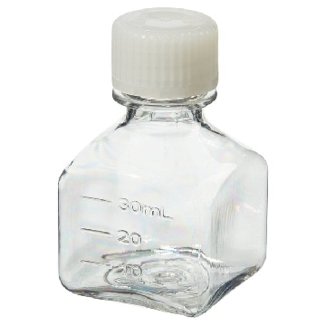
¥ 10277.12
-
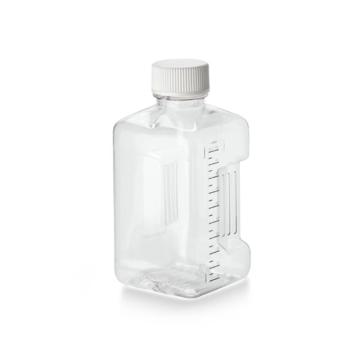
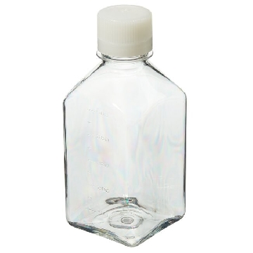 无菌,PETG 方形有刻度培养基瓶,带盖,聚对苯二酸乙二醇酯共聚物,天然高密度聚乙烯盖,500毫升容量,每箱40,40/箱,342020-0500,Nalgene,Thermofisher,赛默飞世尔
¥ 3684.34
-
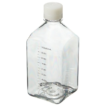
¥ 4404.38
-
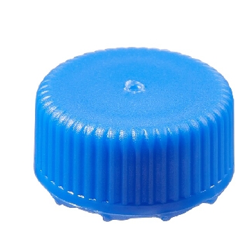
¥ 2617.87









 微信小程序
7X24小时在线咨询
微信小程序
7X24小时在线咨询