

- 商品介绍
- 规格参数
- 包装参数
|
规格 |
|
| Product Line: | PROLONG™ |
| Reagent Type: | Anti-Fade Solution |
| Shipping Condition: | Approved for shipment on Wet or Dry Ice |
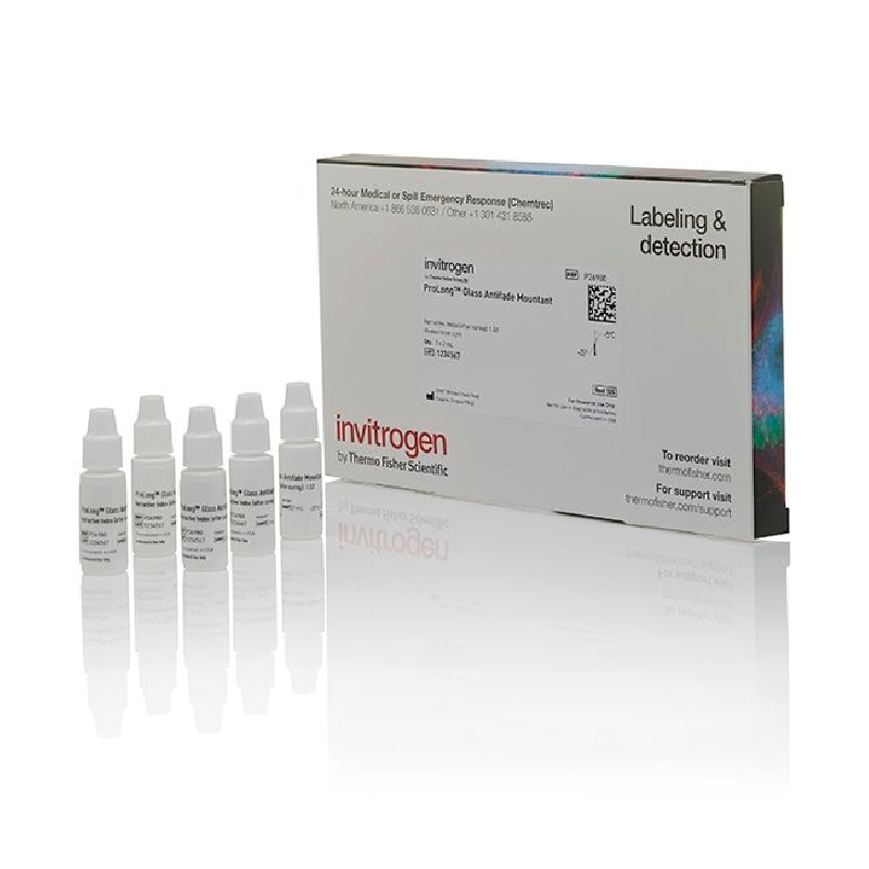
| Quantity: | 5 x 2 mL |
|
储存 |
|
| Store at 2–8°C. For long-term storage, store at -5 to -30°C. Avoid freeze/thaw cycles. |
|
描述 |
ProLong Glass Antifade Mountant is a glycerol-based, hard-setting, ready-to-use mountant that can be applied directly to fluorescently labeled cells or tissue samples on microscope slides or coverslips. It is formulated without DNA stain or with the blue DNA stain NucBlue (Hoechst 33342) or with the deep red DNA stain SYTOX Deep Red. Prolong Glass mountant has a refractive index of 1.52 after curing, similar to that of glass coverslips, compatible immersion oil, and oil-immersion microscope optics, enabling superior resolution and sensitivity. ProLong Glass mountant is also designed to provide unparalleled photobleach protection across the visible and near infra-red spectra. You can use ProLong Glass Antifade Mountant with almost any fluorescent dye or fluorescent protein (e.g., GFP, RFP, mCherry) and with any cell/tissue type for image depths ranging from 0 to 100 µm for bright, high-resolution Z-Stack, 3D, and 2D images. For a non-curing mountant with 1.52 refractive index, try SlowFade Glass Soft-set Antifade Mountant.
With its high refractive index of 1.52 and superior antifade formulation, ProLong Glass Antifade Mountant delivers the following benefits:
• Up to 75% improvement in axial resolution for sharp images
• 3–4 times more imageable focal depth compared tomountants with refractive index of 1.47, with sharp images for monolayers of cells, tissue slices, or 3D cell cultures up to 150 μm thickness/focal depth
• Low background across the spectrum
• Preserved signals from inhibited photobleaching
• Weeks to months of stability for slide-mounted samples
• Ease of use
• Availability with or without DAPI
Learn more about ProLong Antifade mountants >
ProLong Glass Hard-set Antifade Mountant comes already mixed and ready to use in a dropper bottle or 10-mL bottle with or without DNA stain. NucBlue (Hoechst 33342) is excited by UV light at 360 nm when bound to DNA, with an emission maximum at 460 nm, and is detected using a DAPI traditional filter. SYTOX Deep Red stain is excited by red light at 660 nm when bound to DNA, with an emission maximum at 682 nm, and is detected using a Cy5/deep red traditional filter. After removing excess water, just apply a drop of mountant to the sample, add a coverslip, cure, and image. ProLong Glass mountant is a hard-setting, curing mountant that minimizes optical refraction and spherical aberration in the optical path, enables longer-term storage of your samples, and helps protect your samples from photobleaching. ProLong Glass mountant does not discolor or shrink when cured, making it possible to take high-quality fluorescent images weeks, or even months, after mounting the slides.
ProLong Glass Antifade Mountant with NucBlue stain contains an optimized concentration of Hoechst 33342 stain, which provides superior nuclear labeling of most cells and tissues. ProLong Glass mountant with NucBlue stain is not recommended for specimens thicker than 30 µm. Thicker specimens can require longer incubations with labeling reagents, so a pre-incubation with NucBlue Live ReadyProbes Reagent is recomended.
Brightest and sharpest images
Studies show that the axial resolution of a microscopy system can be improved by matching the refractive index of the mounting medium, which maximizes light collection of the objective lens (1–3). Fouquet et. al. saw dramatic improvement in axial resolution while using a confocal microscope with high refractive index media (3). When Prolong Glass Antifade Mountant, which has a refractive index of 1.52, is compared with a mounting medium with a refractive index of 1.47, the axial resolution is improved by 32% at a focal depth of 40 µm and 75% at a focal depth of 100 µm (see figure below). The signal intensity and sensitivity of fluorophores is also improved as the focal depth increases within the sample. The same or greater improvement in resolution and sensitivity is seen in the lateral direction. This makes Prolong Glass a superior mountant for acquisition of Z-Stack, 3D, and 2D images of cells and tissue at depths from 0 to 100 µm (see figure below).
Inhibit photobleaching across the visible and near-IR spectra
No matter which fluorescent dyes or fluorescent proteins you are using, ProLong Glass Antifade Mountant helps provide superior protection against photobleaching. It has been shown to have best-in-class performance with a wide array of fluorescent dyes, including Alexa Fluor, FITC, TRTC, Texas Red, Cy3, and Cy5 dyes (see figure below). ProLong Glass mountant does not quench fluorophores, and its ability to provide brighter signal against no background helps ensure that you get the best-quality high-resolution images for publishing.
Stable signals during imaging and for longer-term storage
ProLong Glass Antifade Mountant is the ideal option for archiving samples with fluorescent dyes or fluorescent proteins and for preserving signal strength during prolonged imaging sessions, such as repeated scanning under confocal laser scanning illumination or with super-resolution microscopy. And it's easy to use—simply remove excess water from the stained sample and mount.
Achieve excellent refractive index matching for crisp images
For modern high-resolution imaging applications, refractive index matching is critical. ProLong Glass Antifade Mountant cures to a refractive index of 1.52. Additionally, this curing property eliminates the need to use messy acrylic resins (i.e., fingernail polish), which can damage or ruin modern microscope objectives.
References
1: Cremer C, Masters BR. Resolution enhancement techniques in microscopy. Eur Phys J. 2013 38(3):281-344.
2: Shimi T, Kittisopikul M, Tran J, Goldman AE, Adam SA, Zheng Y, Jaqaman K, Goldman RD. Structural organization of nuclear lamins A, C, B1, and B2 revealed by superresolution microscopy. Mol Biol Cell. 2015 26(22):4075-86.
3: Fouquet C, Gilles JF, Heck N, Dos Santos M, Schwartzmann R, Cannaya V, Morel MP, Davidson RS, Trembleau A, Bolte S. Improving axial resolution in confocal microscopy with new high refractive index mounting media. PLoS One. 2015 10(3):e0121096.
| 长度(mm) | |
| 宽度(mm) | |
| 高度(mm) | |
| 重量(kg) |
-

询价
-

¥ 1056.74
-

询价
-

询价
-

询价
-

询价
-

¥ 145.30
-
,125条/10包/箱,PCR-0208-C,Axygen,爱思进")
¥ 1039.20
-

¥ 1292.55
-

¥ 3412.00
-
¥ 2369.85
-

询价
-
¥ 7706.18
-

¥ 433.13
-
¥ 3055.44
-

¥ 1509.78
-
¥ 1900.52
-

¥ 879.11
-

¥ 805.95
-
询价


 微信小程序
7X24小时在线咨询
微信小程序
7X24小时在线咨询